

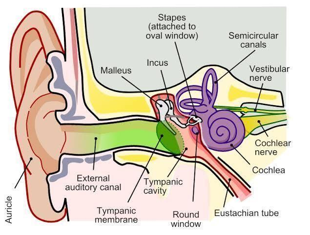
Overview of the Human Ear
The human ear is a remarkable sense organ responsible for two vital functions: hearing and balance. By converting sound waves into nerve impulses, our ears allow us to interpret the sounds around us. Simultaneously, specialised structures within the ear help maintain our body’s equilibrium. In this article, we will discuss a human ear diagram, its easy labelling, the detailed structure of each part, and how these components work together to perform essential functions.
Parts of the Outer Ear
When we think of the parts of the outer ear, the pinna (or auricle) is the most visible. It is shaped to funnel sound waves into the ear canal. Below are the main components of the external ear:
Pinna (Auricle)
The external, flap-like structure collects sound waves from the environment.
Helps in determining the direction from which sounds originate.
External Auditory Canal
A tube-like passage that channels sound waves from the pinna to the tympanic membrane (eardrum).
Lined with tiny hairs and glands that produce earwax (cerumen) to protect the canal.
Tympanic Membrane (Eardrum)
A thin membrane that vibrates when hit by sound waves.
Marks the boundary between the outer ear and the middle ear.
By learning the parts of the outer ear, students can better visualise how sound enters our ears and reaches deeper sections.
Middle Ear
The middle ear amplifies and transmits the vibrations received from the tympanic membrane. It consists of:
Ossicles
Three tiny bones known as the malleus (hammer), incus (anvil), and stapes (stirrup).
These bones are interconnected and act like levers to increase and pass on the vibrations towards the inner ear.
Eustachian Tube
Connects the middle ear to the back of the nose and throat (nasopharynx).
Equalises air pressure on both sides of the tympanic membrane, ensuring accurate sound transmission.
Inner Ear
Often regarded as the most complex part, the inner ear is where sound waves are converted into nerve signals, and where the body’s balance mechanism resides. It comprises:
Cochlea
A coiled, fluid-filled structure containing specialised hair cells (the receptors of hearing).
Transforms mechanical sound vibrations into electrochemical signals sent to the brain via the auditory nerve.
Semicircular Canals
Three loop-shaped canals are positioned at right angles to each other.
Contains fluid and sensory receptors that help detect rotational movements of the head, thus aiding balance.
Vestibule
The central part of the inner ear includes the utricle and saccule.
Senses linear movements and gravitational forces, contributing to balance and spatial orientation.
Human Ear Diagram Labelled
Below is a human ear diagram labelled to help you visualise the outer ear, middle ear, and inner ear. For clarity, you can use different shades to create a human ear diagram with colour. This approach ensures an easy distinction of each part:
Outer Ear: Pinna, External Auditory Canal, Tympanic Membrane
Middle Ear: Ossicles (Malleus, Incus, Stapes), Eustachian Tube
Inner Ear: Cochlea, Semicircular Canals, Vestibule
Human Ear Structure and Function
Understanding the human ear structure and function is crucial for grasping how hearing and balance mechanisms operate:
Hearing: Sound waves enter through the outer ear, causing the tympanic membrane to vibrate. Ossicles in the middle ear amplify these vibrations and pass them on to the cochlea in the inner ear. Sensory hair cells in the cochlea convert these vibrations into nerve impulses that travel to the brain, resulting in the perception of sound.
Balance: The semicircular canals, vestibule, and the fluid within them sense different head movements and positions. They send signals to the brain, enabling us to maintain posture and equilibrium.
Key Highlights
The human ear diagram is typically divided into three main sections: outer ear, middle ear, and inner ear.
The outer ear collects sound waves, the middle ear amplifies them, and the inner ear converts them into electrical signals while also managing balance.
Drawing a human ear diagram easily with proper labels and colour coding helps memorise each part’s role.
The parts of the outer ear include the pinna, external auditory canal, and tympanic membrane.
The middle ear’s ossicles (malleus, incus, stapes) are the smallest bones in the human body.
The cochlea, semicircular canals, and vestibule form the key structures for hearing and balance in the inner ear.
Additional Learning Aids
Mnemonic for Ear Bones
A simple mnemonic for remembering the order of the ossicles from the tympanic membrane inwards is:
My Inner Spaces
M: Malleus
I: Incus
S: Stapes
Quick Quiz
Name the three tiny bones of the middle ear.
What is the function of the Eustachian tube?
How do the semicircular canals help in balance?
Which part of the inner ear detects linear movements of the head?
Answer these questions to test your knowledge of the human ear diagram.
Short Activity
Draw a human ear diagram with colour labelling each part clearly.
Use 3-4 colours to differentiate the outer, middle, and inner ear for easy reference.
Write down one key function beside each labelled part to revise faster.


FAQs on Human Ear Diagram Labelled and Detailed Structure
1. What are the three main sections of the human ear and their primary roles?
The human ear is divided into three main sections: the outer ear, which collects sound waves; the middle ear, which amplifies these sound waves; and the inner ear, which converts the waves into nerve signals that the brain can understand.
2. What is the function of the eardrum in the process of hearing?
The eardrum, or tympanic membrane, is a thin piece of tissue that separates the outer ear from the middle ear. When sound waves travel down the ear canal and hit it, the eardrum vibrates. These vibrations are then passed on to the tiny bones in the middle ear.
3. How does the middle ear make sounds louder?
The middle ear amplifies sound using a set of three tiny, connected bones called ossicles (malleus, incus, and stapes). They act like a lever system. The vibrations from the large eardrum are concentrated onto the much smaller stapes, increasing the pressure and making the sound much stronger before it enters the inner ear.
4. What are the two different jobs performed by the inner ear?
The inner ear is responsible for two different but equally important functions:
- Hearing: The snail-shaped cochlea translates sound vibrations into electrical impulses, which are sent to the brain via the auditory nerve.
- Balance: The vestibular system (including the semicircular canals) detects the movement and position of your head, sending signals to the brain to help you maintain balance.
5. Why is the cochlea shaped like a spiral instead of being a straight tube?
The spiral shape of the cochlea is a highly efficient way to fit a long structure into a very small, bony space in the skull. This length is crucial because different parts of the cochlea are tuned to detect different sound frequencies, from high pitch to low pitch, allowing us to hear a wide range of sounds.
6. How does the ear help us maintain balance when we move our head?
Inside the inner ear, the semicircular canals are filled with fluid. When you move your head, this fluid sloshes around and stimulates tiny hair cells. These cells send signals to your brain about the direction and speed of your movement, allowing your body to adjust and stay balanced.
7. What is the Eustachian tube and why is it important?
The Eustachian tube is a small passageway that connects the middle ear to the back of the throat. Its main job is to equalise the air pressure on both sides of the eardrum. This is why swallowing or yawning can help your ears 'pop' and feel better during a flight or a change in altitude.
8. What are the most important parts to label in a human ear diagram for a school exam?
For most school-level exams, you should focus on being able to identify and label these key structures:
- Outer Ear: Pinna and Auditory Canal.
- Boundary: Eardrum (Tympanic Membrane).
- Middle Ear: The three ossicles (Malleus, Incus, Stapes).
- Inner Ear: Cochlea, Semicircular Canals, and the Auditory Nerve.


