

Downloadable Dicot Leaf Diagram PDF with Clear Labels
The concept of dicot leaf diagram is essential in biology and helps explain real-world biological processes and exam-level questions effectively.
Understanding Dicot Leaf Diagram
Dicot leaf diagram refers to a detailed, labelled illustration showing the internal cross-sectional structure of the leaf of dicotyledonous plants. This concept is important in areas like plant anatomy, practical biology, and board exam preparation. Diagrams help students recognize tissues such as mesophyll, vascular bundles (veins and midrib), epidermis, and stomata, supporting topics like photosynthesis and tissue differentiation.
Structure of Dicot Leaf Diagram
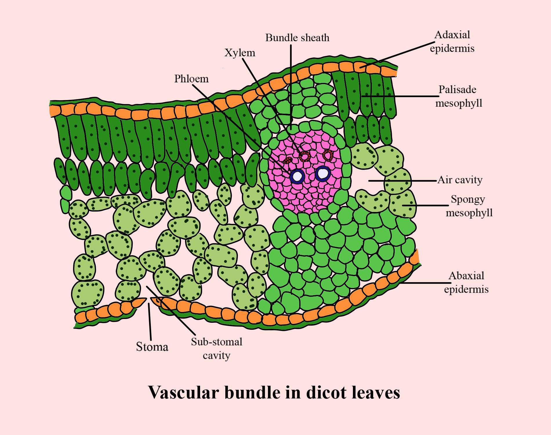
A typical dicot leaf diagram displays three main regions:
- Upper and Lower Epidermis: Thin layers covering the leaf's upper (adaxial) and lower (abaxial) surfaces, sometimes with a waxy cuticle for protection against water loss and injury.
- Mesophyll: Layer between the epidermises, divided into two parts: palisade parenchyma (elongated cells rich in chloroplasts, just below the upper epidermis) and spongy parenchyma (loosely arranged cells with air cavities, near the lower epidermis).
- Vascular Bundles: Comprised of xylem and phloem for water/mineral and food transport; bundled together in the veins and midrib, surrounded by bundle sheath cells for protection.
The lower surface typically has more stomata, important for gas exchange and transpiration.
Key Parts of a Dicot Leaf (Label and Function)
| Part | Location | Function |
|---|---|---|
| Upper Epidermis | Outer upper layer | Protection, sometimes covered with cuticle |
| Palisade Parenchyma | Below upper epidermis | Photosynthesis (rich in chloroplasts) |
| Spongy Parenchyma | Below palisade, above lower epidermis | Air exchange, minor photosynthesis |
| Vascular Bundles | Veins/Midrib | Transport of water, minerals, food |
| Lower Epidermis | Outer lower layer | Has more stomata for gas exchange |
| Stomata | Mainly in lower epidermis | Controls water loss and gas exchange |
Identification Tips and Monocot vs Dicot Leaf
- Mesophyll differentiation: Only dicot leaves have both palisade and spongy mesophyll. Monocots have undifferentiated mesophyll.
- Number of stomata: Dicots have more stomata on lower surface; monocots have equal numbers on both sides.
- Venation: Dicots: reticulate (net-like) venation. Monocots: parallel venation.
- Bundle sheath cells: Well-developed in dicots.
For a detailed difference, visit Difference Between Monocot and Dicot Leaf.
Worked Example – Observing Dicot Leaf Under Microscope
1. Take a section of the leaf using a blade.
2. Place it on a glass slide with water and cover-slip.
3. Observe the arrangement: upper epidermis, palisade cells, spongy mesophyll, vascular bundles, and lower epidermis.
4. Draw and neatly label all visible tissues as shown above.
Tip: Always note the side with more stomata as the lower surface.
Common Mistakes to Avoid
- Confusing dicot leaf diagram with monocot (look at mesophyll and venation).
- Mixing label positions for upper/lower epidermis or forgetting to show bundle sheath.
Real-World Applications
The concept of dicot leaf diagram is used in fields like botany, agriculture, and environmental biology. It is vital for understanding photosynthesis and plant nutrition. In practical labs and exams, drawing and identifying the correct tissues can help score well. Vedantu brings such concepts alive with high-quality visuals and stepwise guidance.
Practice Questions
- Draw and label a neat diagram of the cross-section of a dicot leaf.
- Describe the roles of palisade and spongy parenchyma in the dicot leaf.
- List three anatomical differences between dicot and monocot leaves.
- Why are more stomata present on the lower surface of a dicot leaf?
In this article, we explored dicot leaf diagram, its key parts, structure, practical significance, and tips for identifying it. Practicing labelled diagrams and understanding the roles of tissues builds exam confidence. To learn more, keep practicing with Vedantu.
Related Concepts & Useful Vedantu Links
- Difference Between Monocot and Dicot Leaf
- Plant Cell
- Photosynthesis Process
- Difference Between Monocot and Dicot Root
- Lamina of a Plant Leaf
- Stomata
- Cell Structure and Function
- Nutrition in Plants
- Plant Tissues
- Basic Internal Anatomy of Leaf
- Difference Between Monocot and Dicot Stem


FAQs on Dicot Leaf Diagram: Labeled Structure & Easy Parts
1. What is a dicot leaf diagram?
A dicot leaf diagram is a labeled illustration showing the typical internal structure of a dicotyledonous leaf. It includes important parts such as the upper and lower epidermis, mesophyll (palisade and spongy parenchyma), vascular bundles, and stomata, helping students visualize leaf anatomy for exams and practicals.
2. How do you identify a dicot leaf under a microscope?
To identify a dicot leaf under a microscope, look for a dorsiventral structure with distinct upper (adaxial) and lower (abaxial) surfaces. The palisade parenchyma is present just below the upper epidermis and consists of elongated cells rich in chloroplasts, while the spongy parenchyma beneath has loosely arranged round cells with air spaces. Additionally, more stomata are found on the abaxial surface, and the vascular bundles (containing xylem and phloem) are clearly visible within the midrib and veins.
3. What are the main parts labeled in a dicot leaf diagram?
The main parts labeled in a dicot leaf diagram include:
- Upper Epidermis: Protective outer layer with thin cuticle.
- Palisade Parenchyma: Elongated cells rich in chloroplasts for photosynthesis.
- Spongy Parenchyma: Loosely arranged cells with air spaces for gas exchange.
- Vascular Bundles: Contain xylem for water transport and phloem for food transport.
- Lower Epidermis: Contains more stomata for transpiration and gas exchange.
- Stomata: Pores regulated by guard cells for gas exchange.
4. How is a dicot leaf diagram important for class 10 and 11 exams?
A dicot leaf diagram is essential for class 10 and 11 biology exams because it helps students:
- Clearly understand leaf anatomy and functions.
- Answer practical questions with accurate labeled diagrams.
- Distinguish differences between monocot and dicot leaves.
- Score well in board exams, NEET, and other competitive tests.
- Develop skills in biological drawing and observation.
5. What are the 5 key differences between monocot and dicot leaves?
The key differences between monocot and dicot leaves are:
- Veins: Dicots have reticulate venation, monocots have parallel venation.
- Mesophyll: Dicots show distinct palisade and spongy parenchyma, monocots have undifferentiated mesophyll.
- Stomata Distribution: Dicots usually have more stomata on the lower epidermis; monocots have them evenly distributed.
- Vein Ends: Dicots' veins end freely; monocot veins may anastomose.
- Vascular Bundle Arrangement: Dicots have vascular bundles arranged in rings within the leaf; monocots have scattered bundles.
6. Where can I download a simple, labeled dicot leaf diagram PDF?
You can download a simple, labeled dicot leaf diagram PDF from trusted educational sites like Vedantu and NCERT portals. These PDFs usually include labeled diagrams and practice sheets to aid in revision and exam preparation. For example, Vedantu often provides downloadable resources directly linked on their biology topic pages.
7. Why do students confuse dicot leaf diagrams with monocot leaf diagrams in practicals?
Students often confuse dicot and monocot leaf diagrams because both share some common features at first glance. The confusion arises mainly due to:
- Similar overall leaf shape in some specimens.
- Lack of clear understanding of venation types: reticulate vs parallel.
- Not recognizing the distinction in mesophyll structures.
- Insufficient practice in identifying stomata distribution.
Clear labeling and knowledge of key differences improve accuracy.
8. Which mistakes lower marks when drawing a dicot leaf diagram in boards?
Common mistakes that lower marks include:
- Incorrect or missing labels of major parts like palisade parenchyma or vascular bundles.
- Inaccurate shape or proportion of the leaf and its parts.
- Lack of clarity in showing stomata or epidermis layers.
- Messy, unclear drawing that lacks neatness.
- Not following the prescribed diagram style or syllabus guidelines.
9. How to quickly revise all parts of a dicot leaf without mixing labels?
To quickly revise all parts:
- Use a clear labeled diagram as a visual aid.
- Create mnemonic devices for parts like epidermis, mesophyll, vascular bundles.
- Practice drawing the diagram repeatedly with labels.
- Use downloadable PDFs for on-the-go revision.
- Focus on understanding rather than rote memorization to avoid mixing labels.
10. Why are stomata locations important in a dicot leaf drawing?
The location of stomata is important because it reflects the leaf’s adaptation to gas exchange and transpiration. In a dicot leaf, stomata are primarily found on the abaxial (lower epidermis) surface to minimize water loss while allowing efficient gas exchange. Correctly showing stomata location in diagrams demonstrates a student's understanding of leaf function and anatomy.
11. When is a cross-section view required versus top view in diagrams?
A cross-section view is used to show the internal structure of the leaf, including the arrangement of epidermis, mesophyll, vascular bundles, and stomata. It is essential for detailed understanding and practical exams. The top view or surface view focuses on venation patterns and overall shape, useful for identification and comparison. Exams often require the cross-section view to test detailed anatomy knowledge.


